|
|

■Simple operation for accurate diagnostic information
As eight sensors, which are placed at 6-mm intervals along the catheter for this system, cover the entire measurement area, there is no need to shift its position once the catheter is inserted.
Simply place it, and you will get a measurement more accurate than ever.
■Pressure measurement across the entire anorectal area
This system is capable of measuring both the anal and rectal regions simultaneously to assess the anal pressure distribution, squeezing pressure distribution, the relaxation or its dysfunction during simulated defecation, enabling an easy but detailed assessment of abnormal pressure distributions often seen in patients with postsurgical sphincteric disorders or outlet syndrome.
■Easy-to-grasp Visual Images
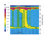
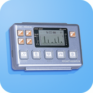
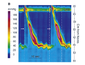
Our pressure measurement system incorporating the PocketMonitor GMS-4000 model displays the status of contraction and relaxation represented in the form of consecutive color topography plots.
In this way, you can visually grasp the function of target organs, which was hardly seen on displayed waveforms only.
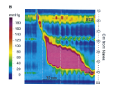
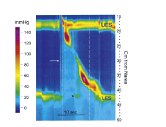
Examples of acquired pressure data measured with pressure transducers
|
Color contour plot showing normal water swallow |
Symptom No.3 |
|
Symptom No.1 |
Symptom No.2 |